home libri books Fumetti ebook dvd top ten sconti 0 Carrello


Torna Indietro

russo paolo (curatore) - handbook of x-ray imaging

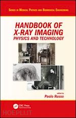
Handbook of X-ray Imaging Physics and Technology


Disponibilità: Normalmente disponibile in 20 giorni
A causa di problematiche nell'approvvigionamento legate alla Brexit sono possibili ritardi nelle consegne.


PREZZO
611,98 €
NICEPRICE
581,38 €
SCONTO
5%


Questo prodotto usufruisce delle SPEDIZIONI GRATIS
selezionando l'opzione Corriere Veloce in fase di ordine.


Pagabile anche con Carta della cultura giovani e del merito, 18App Bonus Cultura e Carta del Docente


Facebook Twitter Aggiungi commento


Spese Gratis

Dettagli

Genere:Libro
Lingua: Inglese
Editore:

CRC Press

Pubblicazione: 01/2018
Edizione: 1° edizione


Note Editore

Containing chapter contributions from over 130 experts, this unique publication is the first handbook dedicated to the physics and technology of X-ray imaging, offering extensive coverage of the field. This highly comprehensive work is edited by one of the world’s leading experts in X-ray imaging physics and technology and has been created with guidance from a Scientific Board containing respected and renowned scientists from around the world. The book's scope includes 2D and 3D X-ray imaging techniques from soft-X-ray to megavoltage energies, including computed tomography, fluoroscopy, dental imaging and small animal imaging, with several chapters dedicated to breast imaging techniques. 2D and 3D industrial imaging is incorporated, including imaging of artworks. Specific attention is dedicated to techniques of phase contrast X-ray imaging. The approach undertaken is one that illustrates the theory as well as the techniques and the devices routinely used in the various fields. Computational aspects are fully covered, including 3D reconstruction algorithms, hard/software phantoms, and computer-aided diagnosis. Theories of image quality are fully illustrated. Historical, radioprotection, radiation dosimetry, quality assurance and educational aspects are also covered. This handbook will be suitable for a very broad audience, including graduate students in medical physics and biomedical engineering; medical physics residents; radiographers; physicists and engineers in the field of imaging and non-destructive industrial testing using X-rays; and scientists interested in understanding and using X-ray imaging techniques. The handbook's editor, Dr. Paolo Russo, has over 30 years’ experience in the academic teaching of medical physics and X-ray imaging research. He has authored several book chapters in the field of X-ray imaging, is Editor-in-Chief of an international scientific journal in medical physics, and has responsibilities in the publication committees of international scientific organizations in medical physics. Features: Comprehensive coverage of the use of X-rays both in medical radiology and industrial testing The first handbook published to be dedicated to the physics and technology of X-rays Handbook edited by world authority, with contributions from experts in each field


Sommario

PART 1: Basic physical and technological aspects Chapter 1: Basic physics of X-ray interactions in matter Chapter 2: Xray tube physics and technology Chapter 3: X-ray generators Chapter 4: Carbon Nanotube Based Field Emission X-ray Technology Chapter 5: Technology of Miniature X-Ray Tubes Chapter 6: Technology of pyroelectric X-ray tubes Chapter 7: History of X-Ray Tubes Chapter 8: Synchrotron radiation x-ray sources for radiography and tomography Chapter 9: X-ray shutters Chapter 10: Calculation of x-ray spectra Chapter 11: Radiochromic film dosimetry for radiology Chapter 12: Computed Radiography Chapter 13: Photon Counting Detectors for X-ray imaging Chapter 14: Image Quality Chapter 15: Image quality in attenuation based and phase contrast based X-ray imaging Chapter 16: Inverse Compton Scattering X-ray sources PART 2: X-ray Radiography and Fluoroscopy Chapter 17: Wilhelm Conrad Roentgen - The discovery of X-rays and the creation of a new medical profession Chapter 18: History of Radiology Chapter 19: Digital Mammography Chapter 20: Digital Breast Tomosynthesis Chapter 21: Fluoroscopy: physics and technology Chapter 22: Dental radiography Chapter 23: Clinical mammographic and tomosynthesis units Chapter 24: Physical image quality evaluation of x-ray detectors for digital radiography and mammography Chapter 25: Mammography, breast tomosynthesis and risk of radiation-induced breast cancer Chapter 26: Clinical radiographic units Chapter 27: Clinical Fluoroscopy units Chapter 28: Physical Basis of X-ray Breast Imaging Chapter 29: Radiation dose in X-ray mammography and digital breast tomosynthesis Chapter 30: Industrial Radiography Chapter 31: Forensic Radiology PART 3: X-ray Computed Tomography Chapter 32: X-ray Computed Tomography for diagnostic imaging – from single-slice to multi-slice Chapter 33: Analytical reconstruction methods in X-ray CT Chapter 34: Iterative reconstruction methods in X-ray CT Historical image gallery Chapter 35: X-ray Cone-Beam Computed Tomography Chapter 36: Small Animal X-ray Computed Tomography Chapter 37: Quality Assurance of X-ray Computer Tomography Chapter 38: Radiation Dose in X-ray Computed Tomography Chapter 39: Dual energy X-ray computed tomography Chapter 40: Soft x-ray tomography: techniques and applications Chapter 41: 4-D X-ray Computed Tomography Chapter 42: Dental and Maxillofacial Cone Beam Computed Tomography Chapter 43: High Speed X-ray computed tomography Chapter 44: Kilovoltage and Megavoltage Imaging in Radiotherapy Chapter 45: Industrial X-ray computed tomography Chapter 46: Industrial X-ray computed tomography scanners Chapter 47: Dimensional metrology for industrial Computed Tomography Chapter 48: Influence of scatter in X-ray imaging and scatter correction methods for industrial applications PART 4: Phase-contrast X-ray imaging and other aspects Chapter 49: Theory of X-ray phase-contrast imaging Chapter 50: Non-interferometric techniques for X-ray phase-contrast biomedical imaging Chapter 51: X-ray phase-contrast mammography Chapter 52: X-ray phase contrast tomosynthesis imaging Chapter 53: Crystal analyser-based X-ray Phase Contrast Imaging Chapter 54: X-Ray scattering: analytical applications and imaging Chapter 55: Tissue Substitute Materials for Diagnostic X-ray Imaging Chapter 56: Phantoms for image quality and dose assessment Chapter 57: Software phantoms for X-ray radiography and tomography Chapter 58: Radiography and Computed Tomography for Works of Art Chapter 59: Computer Aided Diagnosis for X-ray imaging Chapter 60: Computer analysis of mammograms Chapter 61: Databases for mammography Chapter 62: Computer analysis of CT images for lung nodule detection Chapter 63: Display Optimization & Human Factors Chapter 64: Display for Medical Imaging and DICOM Grayscale Standard Display Function Fundamentals Chapter 65: Quality Control of Medical Imaging Displays Chapter 66: Radiation protection issues in X-ray radiology, fluoroscopy and computed tomography Chapter 67: Educational aspects in radiography physics and technology Chapter 68: Tables of X-rays mass attenuation coefficients, of K and L –energy, of K, L and M fluorescence yield, of Ka/Kß, La/Lßand La/L?


Autore

Paolo Russo was born in Naples, Italy, in 1958. In 1981, he graduated in Physics from the University of Naples Federico II. From 1984, he was a Research Associate at the University before becoming an Associate Professor in 1992 and then a Full Professor of Medical Physics in 2001. His scientific research is focused on the development of medical imaging systems for digital radiography, computed tomography, and nuclear medicine. The applications of such systems includes autoradiography with microstrip and pixel semiconductor detectors; digital mammography with photon counting detectors; small animal SPECT and semiconductor compact gamma cameras; and a prototype scanner for cone-beam breast computed tomography using attenuation based and phase contrast based methods. From 2008 to 2012, Paolo served as the Associate Editor of the journal "Physica Medica" (European Journal of Medical Physics), before being appointed as the Editor-in-Chief of this journal in January 2013. He is also the vice-chair of the Publications Committee of the IOMP and the chair of the Publications and Communications Committee of the EFOMP. In addition, he frequently acts as a reviewer for several journals in Medical Physics and for research projects for the European Research Foundations. Dr. Russo has co-authored over 140 papers in peer reviewed journals and many scientific book chapters.


Altre Informazioni

ISBN:

9781498741521

Condizione: Nuovo
Collana: Series in Medical Physics and Biomedical Engineering
Dimensioni: 11.01 x 8.25 in Ø 7.55 lb
Formato: Copertina rigida
Illustration Notes:1315 b/w images, 136 tables, 685 halftones and 630 line drawings
Pagine Arabe: 1393
Pagine Romane: xxvi


Dicono di noi